
"I notice there is 2" (50.8mm) wafer available on your website and I can go with that. We are using this wafer for oil-immersion microscopy so the thickness under 150um is necessary."
Thin Sapphire Wafers for Oil Immersion Microscopy
UniversityWafer supplies thin sapphire wafers for oil immersion microscopy, fluorescence imaging, confocal microscopy, optical inspection, and laboratory research. Sapphire is a strong choice for microscopy applications because it is transparent, scratch resistant, chemically durable, and available in thin polished formats.
A scientist contacted us looking for a thin sapphire substrate for oil-immersion microscopy:
UniversityWafer, Inc. quoted:
- 50.8mm sapphire wafers
- 100 µm thickness
- Double-side polished (DSP)
- C-plane sapphire
- Off to M-plane 0.2°
Thin sapphire wafers under 150 µm are often used when researchers need a durable transparent substrate for high-resolution imaging. Price depends on diameter, thickness, polish, orientation, and quantity.
Need thin sapphire wafers for microscopy? Buy sapphire wafers online or request a custom quote for your optical imaging application.
What is Oil Immersion Microscopy?
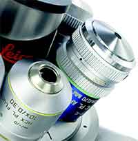 Oil immersion microscopy is a technique used to improve image resolution at high magnification. A drop of immersion oil is placed between the microscope objective and the sample area to reduce light refraction and help more light enter the objective lens.
This method is commonly used with 100x oil immersion objectives for biological imaging, bacteria observation, fluorescence microscopy, live cell imaging, and high-resolution inspection of small features. The oil helps create a clearer optical path between the lens and the sample surface.
Why Use Thin Sapphire Instead of Standard Glass?
Standard glass slides work for many microscopy applications, but sapphire wafers offer stronger durability when the research requires a harder, more chemically resistant, and more scratch-resistant transparent material. Thin sapphire substrates are especially useful for demanding optical setups where surface quality and long-term stability matter.
Sapphire can be useful for:
- Oil immersion microscopy
- Fluorescence microscopy
- Confocal microscopy
- Raman spectroscopy
- Optical imaging research
- Bioimaging substrates
- Semiconductor inspection
Why Use Sapphire Wafers for Oil Immersion Microscopy?
Sapphire wafers are ideal for oil immersion microscopy because they provide excellent optical transparency, high scratch resistance, chemical durability, and mechanical strength. These properties make thin sapphire substrates useful for high-resolution microscopy, fluorescence imaging, confocal microscopy, Raman spectroscopy, semiconductor inspection, and photonics research.
In oil immersion microscopy, immersion oil helps reduce light refraction between the microscope objective and the sample. A thin, double-side polished sapphire wafer can provide a flat, durable, transparent support surface for imaging applications that require optical clarity and long-term substrate stability.
Thin Sapphire Substrates for Optical Imaging
Researchers often request thin sapphire wafers for microscopy when glass slides are not durable enough. Sapphire offers strong optical performance with better hardness, chemical resistance, and thermal stability than many standard glass materials.
Thin sapphire wafers under 150 µm are commonly used for oil immersion microscopy, fluorescence microscopy, bioimaging, optical testing, semiconductor inspection, and other research applications requiring transparent polished substrates.
Common Microscopy Applications
- Oil immersion microscopy
- Fluorescence microscopy
- Confocal microscopy
- Raman spectroscopy
- Bioimaging research
- Semiconductor inspection
- Photonics and optical device research
Sapphire vs. Glass for Microscopy
Sapphire wafers can be a better choice than traditional glass slides when the application requires a harder, more scratch-resistant, chemically stable substrate. Sapphire has a Mohs hardness of 9, which helps protect the surface during handling, cleaning, and repeated laboratory use.
Because sapphire maintains strong optical transparency across a broad wavelength range, it is used in UV imaging, infrared optics, high-precision microscopy, and advanced optical systems. Researchers may also combine sapphire with quartz components, optical coatings, or photonic structures for specialized imaging setups.
Useful Sapphire Wafer Specifications
For microscopy and imaging applications, researchers often choose sapphire wafers based on thickness, diameter, orientation, polish, and surface quality. Double-side polished sapphire wafers are especially useful when light transmission and low optical distortion are important.
- Thin sapphire wafers under 150 µm
- Double-side polished surfaces
- C-plane sapphire orientation
- High optical transparency
- Strong chemical resistance
- Excellent scratch resistance
- Custom diameters and thicknesses available